
Glycans in Health and Disease
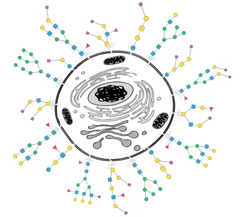
The time has passed when glycans were considered not more than simple decorations of other life-important macromolecules. Meanwhile, there is no doubt that glycans, along with DNA, proteins, and lipids, are essential for all forms of life – from unicellular to multicellular organisms. These ‘’decorations’’, found on the surface of all living cells and nearly all extracellular molecules, are involved in virtually all fundamental molecular processes – ranging from molecular trafficking, clearance, and quality control, through cell-cell recognition and adhesion, to signal transduction and receptor activation. Despite their importance, the structural complexity of glycans, together with associated difficulties in their analysis, make it challenging to obtain exact insights into the roles of glycans in health and disease. Therefore, we strive to develop innovative glycoanalytical tools that can cope with these challenges and that enable in-depth glycosylation analyses. This applies to small amounts of glycans in biological samples, large patient sample cohorts, and samples that harbor multiple classes of glycans. Some of the applications and collaborations in this field are summarized below.

Embryonic development is a biological process that is still not fully understood. This concerns also the role of specific classes of glycans during this process. The tools for glycan and glycopeptide analyses, developed in our lab, help to detect glycosylation changes in sialylation-impaired mouse models and consequently to better understand how sialylation affects early murine embryonic development in vitro [Abeln et al. (2017)]. To gain a deeper understanding of the functional role of N-glycosylation, O-mannosylation, and C-mannosylation on the molecular, cellular and organismal level, we are analyzing in a DFG-funded project the glycosylation of model glycoproteins, patient-derived cell lines, as well as model organisms using a glycomics and glycoproteomics toolbox developed by our group [2]. In particular, with our partners, we focus on the interplay between various ER-initiated protein glycosylation pathways in the context of congenital disorders of glycosylation (CDG).
Cells in the human body are covered with a dense layer of glycans, to which both – "own" and "foreign" cells and proteins can bind. This includes pathogens such as bacteria and viruses, which bear their own unique glycan structures that are important for their life-cycle, i.e. host invasion, immune evasion and pathogen spread. Glycoanalysis methods established by our group were applied to pinpoint producer cell-dependent N-glycosylation differences of simian immunodeficiency virus (SIV) envelope glycoproteins that profoundly impact viral infectivity, viral capture by immune cells lectins, neutralization by antibodies, and mucosal transmission [3].
Because glycan synthesis is not template-driven but depends on the fine interplay of hundreds of genes, it is inherently sensitive to alterations in cellular physiology, i.e. intra- and extracellular stimuli (intracellular: e.g. stage of the cell cycle, mutations; extracellular: e.g. type of nutrition, bacterial infections). Glycans, therefore, can undergo specific changes that can be the cause or the by-product/outcome of a disease. It is now well-established that glycans are directly involved in the pathophysiology of an increasing number of diseases. Thus, investigation of disease-associated changes in glycosylation can be crucial for better understanding of the underlying mechanisms of disease, as well as for diagnosis and therapy. Glycoanalytical tools allow us to profile changes in protein glycosylation associated with diseases such as cancer [4], autoimmune and inflammatory diseases [5,6], as well as congenital disorders of glycosylation [2].
Given the strong correlation of altered glycosylation patterns with many diseases, changes in glycosylation can be crucial in early disease diagnosis, prognosis, and treatment monitoring. Therefore, we started to evaluate the potential of the plasma N-glycome for disease biomarker discovery. A long-term stability study of the plasma N-glycome demonstrated that the individual N-glycome is remarkably stable over several years, while inter-individual differences are enormous. Consequently, taking time-series from individuals enabled to detect small disease-related changes in the N-glycome, improving the diagnostic potential of glyco-biomarkers [7]. This work was continued by two studies investigating cancer-associated changes of the skin N- and O-glycome [4], as well as rheumatoid arthritis-associated changes of the plasma N-glycome during pregnancy [5]. Ongoing work focuses on the explorative investigation of disease-related N-glycosylation alterations of human blood plasma proteins using in-depth glycoproteomic approaches. This endeavor seeks to detect and validate N-glycosylation-based disease-related biomarkers for various auto-immune diseases, including rheumatoid arthritis and inflammatory bowel disease.

